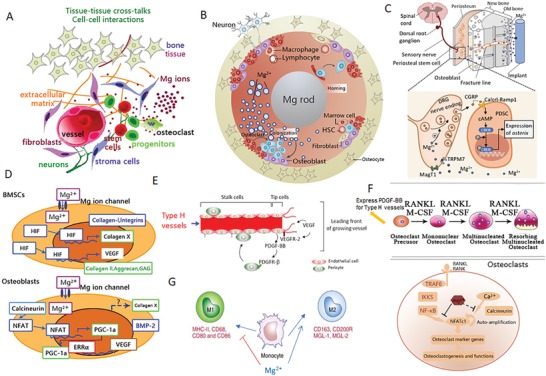
Figure 5.

Cellular and molecular mechanisms demonstrating the potential benefits of Mg ions on bone homeostasis. A) Schematic diagram showing the stimulation of Mg ions (released from Mg‐based implants) on the cross‐talk between connecting tissues (bone, nerves, and vessels), as well as the interactions between cells (stem cells, osteoblast, osteocytes, osteoclasts, endothelial cells, and macrophages); B) Cross‐sectional view of the cellular components affected by the release of Mg ions from an intramedullary orthopaedic implant; C) Robust bone formation at the periosteal region demonstrating the differentiation of periosteum stem cells (PSCs) through the activation of Mg‐induced calcitonin gene‐related peptide (CGRP). This further demonstrates the underlying mechanism that improves healing of osteoporotic bone fractures at the femoral mid‐shaft. Reproduced with permission.18 Copyright 2016, Nature. D) Mg ions directly promote the expression of hypoxia‐induced factor (HIF) in bone marrow mesenchymal stem cells (BMSCs), leading to enhanced chondrogenesis (increased collagen II, aggrecan, and collagen X) and osteogenesis (increased collagen I, BMP‐2, and integrins). Adapted with permission.53 Copyright 2014, Elsevier. E) Mg‐induced production of vascular endothelial growth factor (VEGF) is an essential factor for neo‐formation of type H (CD31hiEndomucinhi) vessels, which may regulate bone homeostasis. Reproduced with permission.129 Copyright 2014, Atlas of Genetics and Cytogenetics in Oncology and Haematology. F) Mg ions inhibit the differentiation of osteoclast precursors by suppressing NF‐κB and NFATc1. High endogenous expression of PDGF‐BB in osteoclast precursors may enhance type H vessel formation. Adapted with permission.52 Copyright 2014, Elsevier. G) Mg ions may promote macrophages to polarize toward the M2 phase that promotes tissue regeneration, instead of M1 phase that promotes an inflammatory response.
