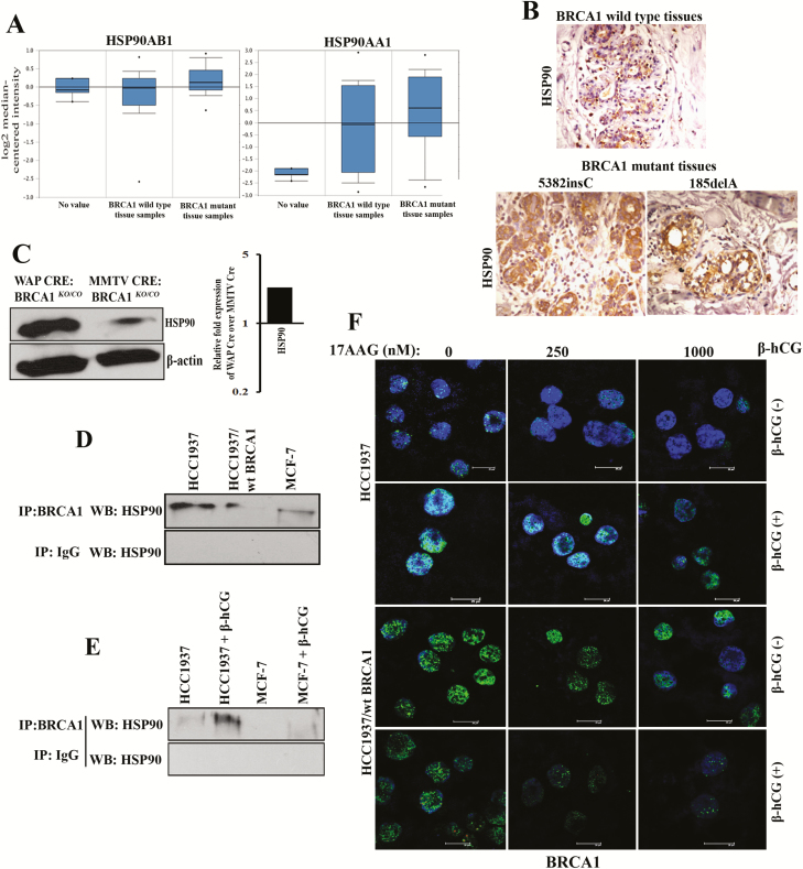
Figure 4.
Presence of β-hCG results in the stabilization of BRCA1 even in the presence of HSP90 inhibitor. (A) TCGA dataset analysis of HSP90AB1 and HSP90AA1 in BRCA1 wild-type and BRCA1 mutant human breast cancer tissue samples (Waddell’s dataset) using Oncomine. (B) Immunohistochemical analysis of HSP90 in BRCA1 wild-type and BRCA1 mutant (5382insC and 185delA) human breast cancer tissues. (C) Western blot analysis of HSP90 from WAP-Cre; BRCA1KO/CO and MMTV-Cre; BRCA1KO/CO mouse tumor samples. Quantitation of the western blot is given on the right panel. (D) Western blot analysis of HSP90 immunoprecipitated with BRCA1 or IgG antibody in HCC1937, HCC1937/wt BRCA1 and MCF-7. (E) Western blot analysis of HSP90 immunoprecipitated with BRCA1 or IgG antibody in HCC1937 and MCF-7 supplemented with β-hCG. (F) Immunofluorescence for BRCA1 upon treating with 17AAG for a period of 24 h in the presence (+) or in the absence (−) of β-hCG in HCC1937 and HCC1937/wt BRCA1. Scale bars, 20 μm. Blue color in the immunofluorescence images represents 4′,6-diamidino-2-phenylindole, which stains the nucleus, whereas the green color indicates the staining for BRCA1 protein. Full blots of cropped images are included in supplementary information.

