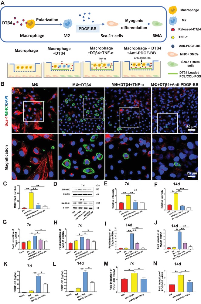
Figure 6.

M2‐Polarized Macrophages promoted myogenic Differentiation Sca‐1+ stem cells by secreting PDGF‐BB. A) Schematic illustration of Sca‐1+ stem cells cocultured with macrophages seeded on nanofibrous scaffolds. B) Immunofluorescent staining of Sca‐1+(red) stem cells, MHC+(green) SMCs, and nuclei (blue) after 7 d coculturing with macrophages in MΦ, MΦ+DTβ4, MΦ+DTβ4+TNF‐α, and MΦ+DTβ4+Anti‐PDGF‐BB groups. C) Comparison of the number of MHC+ cells in MΦ, MΦ+DTβ4, MΦ+DTβ4+TNF‐α, and MΦ+DTβ4+Anti‐PDGF‐BB groups (n = 5 independent samples). D–F) Western blotting reveals SM‐MHC expression in Sca‐1+ stem cells on days 7 and 14 (n = 3 independent samples). G–J) Relative mRNA expressions of Tagln and myh11 in Sca‐1+ cells cocultured with macrophage stimulated by different condition for 7 and 14 d (n = 3 independent samples). Blank: Sca‐1+ cells cultured with unconditioned medium. K,L) Quantification of PDGF‐BB secreted by macrophages in MΦ, MΦ+DTβ4, and MΦ+DTβ4+TNF‐α groups at 7 and 14 d (n = 3 independent samples). M,N) Relative mRNA expressions of PDGF‐BB in macrophages after different treatments for 7 and 14 d (n = 3 independent samples). Blank: macrophages cultured with unconditioned medium. mRNA expression of Tagln, myh11, and PDGF‐BB were analyzed by RT‐qPCR using the ∆∆CT method. Data are represented as the mean ± SD for each group. For (C,E–N), significance was determined by one‐way ANOVA followed by Tukey's post hoc analysis. *: p < 0.05, **: p < 0.01.
