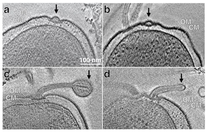
Figure 2.
Cryo-ET reconstructions of intact cells show early stages of flagellar assembly and sheath formation. (a,b) Two representative sections from cryo-ET reconstructions of H. pylori cells show flagellar basal bodies without hook and filament. (c,d) Two representative sections from cryo-ET reconstructions of H. pylori cells show short flagellum. The outer (OM) and cytoplasmic membranes (CM) are indicated.

