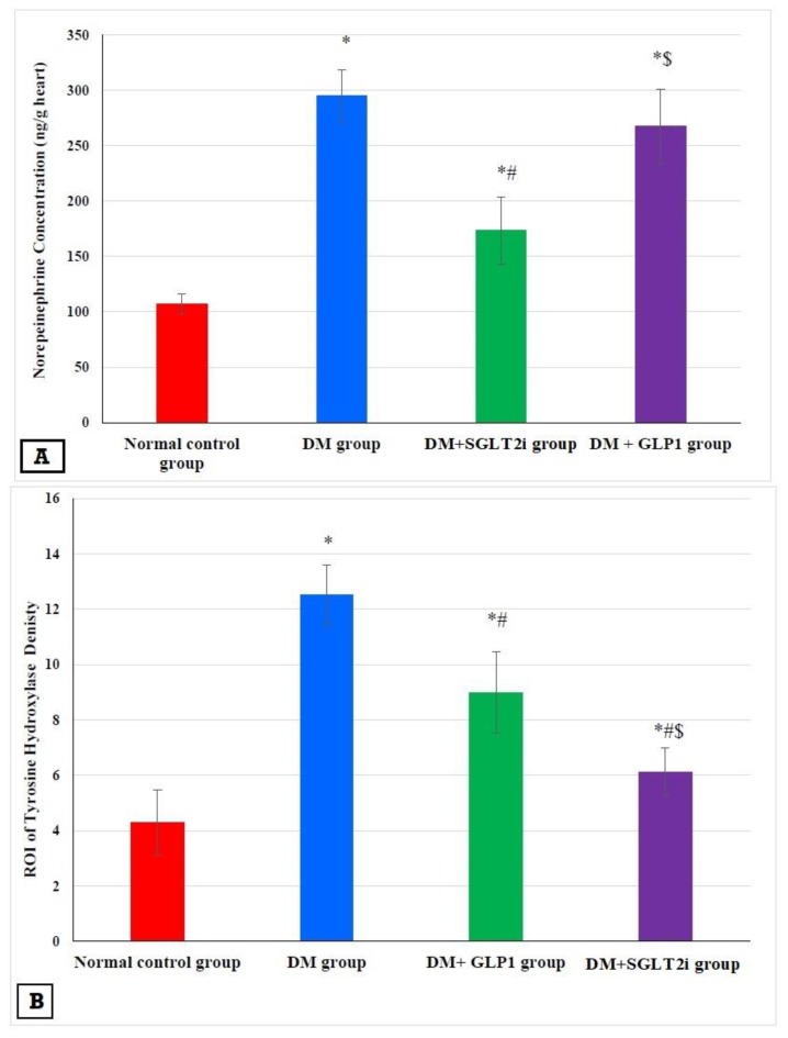
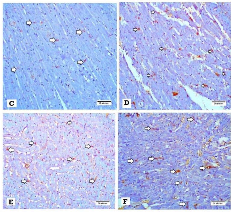
Figure 5.
Effects of SGLTi and GLP1 on the myocardial norepinephrine content and density of sympathetic nerve fibers by tyrosine hydroxylase (TH). (A) The concentration of myocardial NE in different groups, (B) the density of sympathetic nerve fibers by TH immunostaining in different groups, (C) heart specimen from the control group showing evenly distributed TH-positive nerve fibers in the ventricular myocardium (arrows) (400×), (D) heart specimen from the DM group showing marked increase in immunopositivity for TH-positive fibers (arrows) (400×), (E) heart specimen from the DM +GLP1 group showing moderate increase in immunopositivity for TH-positive fibers (arrows) (400×), and (F) heart specimen from the DM +SGLT2i group showing minimal increase in TH-positive fibers (arrows) (400×). * Significant vs. control group, # significant vs. DM group, and $ significant vs. DM + SGLT2i group.


