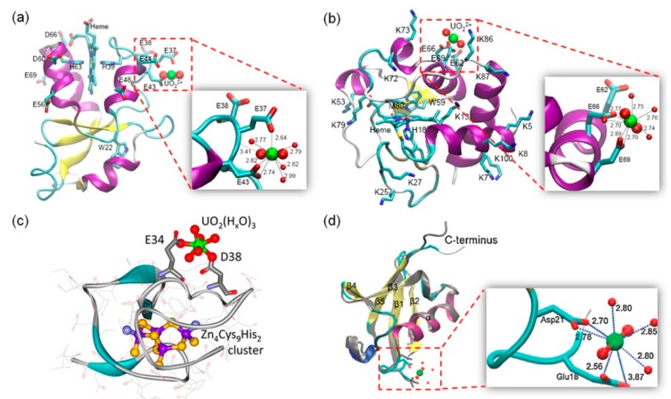
Figure 2.
Modeling structures of UO22+ binding to protein surface. (a) A model of UO22+ binding to Cyt b5 at surface residues, Glu37 and Glu43 [44]; (b) A model of UO22+ binding to Cyt c at surface residues, Glu66 and Glu69 [45]; (c) A model of UO22+ binding to Zn4SmtA species. Reprinted with permission from Ref. [49], Copyright 2016 American Chemical Society; (d) A model of UO22+ binding to Ub at surface residues, Glu18 and Asp21 (cyan). The structure of free Ub (gray) was shown for comparison [53]. Close-up views of the uranyl binding sites were shown as insets, highlighting the coordination and H-bonding interactions.

