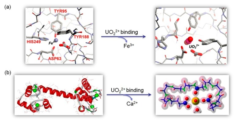
Figure 3.
(a) An X-ray crystal structure (PDB code 1A8E) showing the coordination environment of Fe3+ in the N-lobe of Tf (left), and a proposed model of uranyl binding to the Fe3+ site (right) Reprinted with permission from Ref. [61], Copyright 2007 American Chemical Society. (b) An X-ray crystal structure (PDB code 1EXR) showing the binding of four Ca2+ ions in CaM (left), and a theoretical model of uranyl binding with electronic density based on DFT calculation (right). Reprinted with permission from Ref. [65], Copyright 2016 American Chemical Society.

