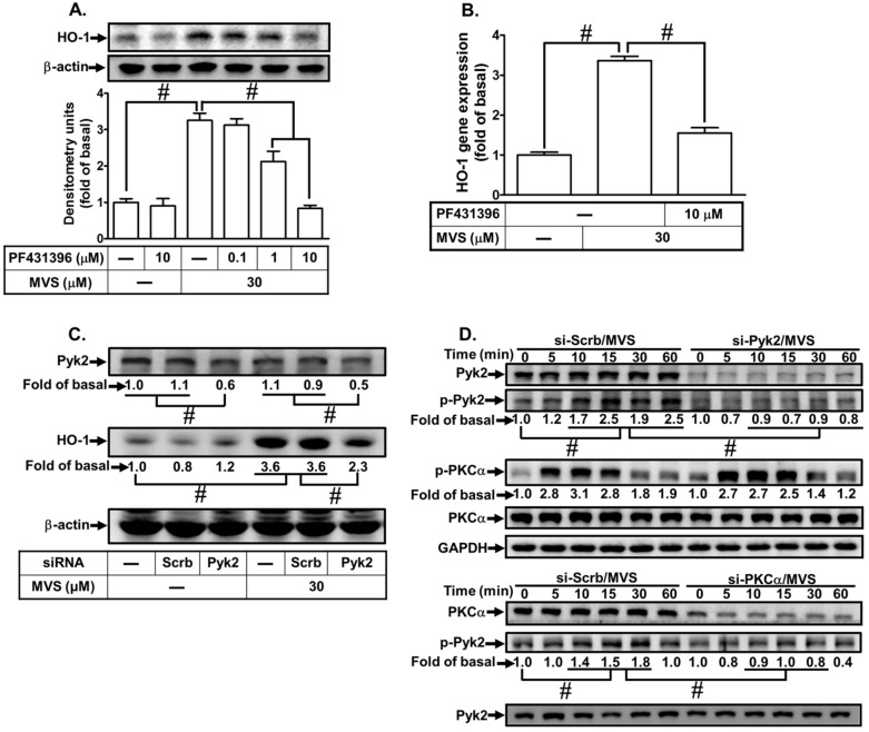
Figure 3.
Phosphorylation of Pyk2 is involved in the MVS-induced HO-1 expression. (A) HPAEpiCs were pretreated with various concentrations of PF431396 for 1 h, and then incubated with vehicle or MVS (30 μM) for 24 h. The levels of HO-1 and β-actin protein expression were determined by Western blot. (B) The cells were pretreated with PF431396 (10 μM) for 1 h and then incubated with vehicle or MVS (30 μM) for 8 h. The levels of HO-1 mRNA were analyzed by real-time qPCR. (C) HPAEpiCs were transfected with scrambled (Scrb) or Pyk2 siRNA, and then incubated with MVS for 24 h. The levels of Pyk2, HO-1, and β-actin protein expression were determined by Western blot. (D) HPAEpiCs were transfected with PKCα or Pyk2 siRNA and then treated with MVS for the indicated time intervals. The levels of phospho-PKCα, PKCα, phospho-Pyk2, Pyk2, and GAPDH were determined by Western blot using respective antibodies. Data are expressed as mean ± SEM of five independent experiments (n = 5). # p < 0.01, as compared with the cells exposed to the indicated reagents.

