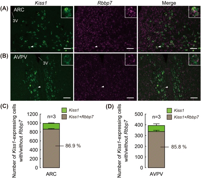
Fig. 2.
Co-expression of Kiss1 and Rbbp7 in the arcuate nucleus (ARC) and anteroventral periventricular nucleus (AVPV) of ovariectomized + low 17β-estradiol (OVX + low E2) rats. Representative photomicrographs showing Kiss1-positive cells (green) and Rbbp7-positive signals (magenta) in the ARC (A) and AVPV (B). The insets indicate expression of Rbbp7-positive signals in the Kiss1-positive cells at a higher magnification (arrowheads). The numbers of Kiss1- and Rbbp7-positive cells out of the Kiss1-positive cells were quantified in the ARC (C) and AVPV (D). Data are the mean ± SEM (n = 3). Scale bars: 100 μm. 3V, third cerebroventricle.

