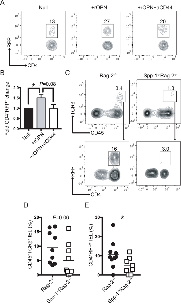
Figure 5.
Osteopontin sustains Foxp3 expression. (A) Intestinal T cells from RFP-Foxp3 reporter mice were isolated and cultured in the presence or absence of recombinant osteopontin (2 μg/ml) and anti-CD44 (5 μg/ml). Seventy-two hours later, the percentage of RFP+ cells was determined by flow cytometry. After excluding dead cells, dot plots were gated as CD45+TCRβ+ cells. (B) Bar graph indicates the fold change in CD4+RFP+ cells in relation to the null group. Data are representative of three independent experiments. (C) Enriched splenic RFP+ cells from RFP-Foxp3 reporter mice were adoptively transferred into Rag-2−/− and Spp-1−/−Rag-2−/− recipient mice. Eight weeks after transfer, IEL from small intestine were isolated and the percentage of RFP+ determined. After excluding dead cells, plots were gated as indicated by the arrows. (D) Summary of the percentage of donor-derived cells recovered; each dot represents an individual mouse. Data are representative from three independent experiments. (E) Summary of the percentage of RFP+ cells within the donor-derived cells. *P<0.05 (Mann-Whitney T test). rOPN = recombinant osteopontin; aCD44 = anti-CD44 antibodies.

