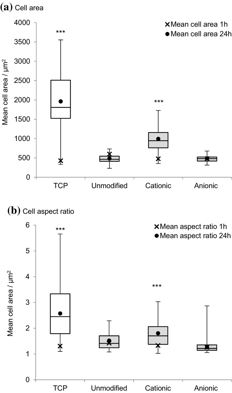
Fig. 10.
Change in MG-63 cell morphology; a cell area and b aspect ratio on cationic, anionic and unmodified bacterial cellulose scaffolds after 1 and 24 h incubation at 37 °C in 5% CO2, n = 17–53 cells measured and error bars show standard error. Cell images were analysed using ImageJ to calculate the average cell aspect ratio and area. The control was tissue culture plastic (TCP). Values significantly different from unmodified cellulose films were indicated by the confidence values *p < 0.05, **p < 0.01 and ***p < 0.001

