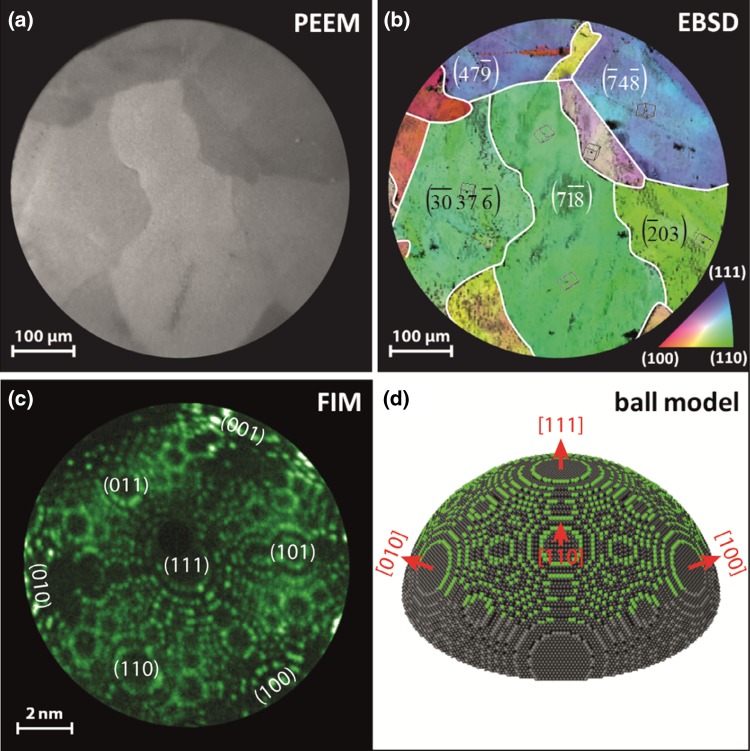
Fig. 1.
Model systems of stepped Rh surfaces a PEEM image of a clean polycrystalline Rh foil surface consisting of μm-sized differently oriented high Miller index domains; b EBSD colorcoded map of the same region with indicated crystallographic orientations of individual domains. The inverse pole figure is shown for reference in the bottom right corner; c Ne + FIM image of the [111]-oriented Rh nanotip; d the 3D ball model based on the atomically resolved FIM images. Atoms visible in the FIM micrograph are marked in green

