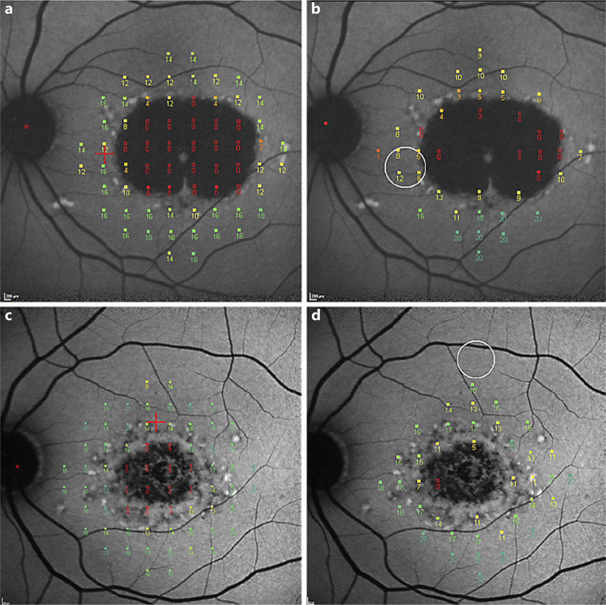
Fig. 2.

Results from mesopic (a, c) and scotopic (b, d) microperimetric exams super-imposed onto the corresponding fundus autofluorescence images of corresponding eyes. The eye shown in a and b shows a lesion of definitely decreased autofluorescence (this type of lesion has at least 90% darkness level compared to the optic nerve head [OHN]). The eye in c and d shows a lesion of questionably decreased autofluorescence (this lesion has darkness levels between 50– and 90% compared to OHN). Sensitivity values for the individual locations (range 0–20 dB) are shown.
