Figure 4.
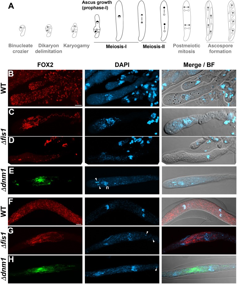
FIS1 and DNM1 elimination affects peroxisome division and distribution during meiocyte differentiation. (A) P. anserina sexual development from the dikaryotic stage to ascospore formation (from left to right, for further details consult Supplementary Figure 1), the developmental stages analyzed in this figure are depicted in black. (B–H) Analysis of FOX2-labeled peroxisomes during meiotic development in WT, Δfis1, and Δdnm1 homozygous crosses. (B–E) Show the peroxisome arrangement in growing meiotic prophase-I asci, and (F–H) in asci at the end of meiosis. Arrowheads point to fragments of non-nuclear genetic material (n, nucleus). Fluorescence images show maximum-intensity projections of z series through the entire cells. Bright field (BF) merged images show single plane micrographs. Scale bar, 5 μm.

