Fig. 2.

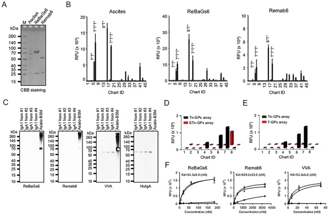
Remab6 is specific for Tn glycopeptides, but not Tn on IgA1, STn, T glycopeptides, BGA or glycans expressing terminal GalNAc. (A) Remab6 and ReBaGs6 were recombinantly expressed in the HEK293 freestyle expression system. The purified Remab6 and ReBaGs6 were separated by SDS-PAGE and stained by CBB solution. (B) Tn glycopeptide (GP) array was probed with ReBaGs6 (middle) and Remab6 (right) to compare to the specificity of the original mouse ascites (left). Chart ID corresponds to Table I. (C) Western blots with IgA1 purified from four individual healthy donor serum (control: Asialo-BSM) were probed with ReBaGs6, Remab6, lectin VVA and goat anti-human IgA antibody. (D–E) Enzymatically remodeled Tn glycopeptide array slides to create STn (D) and T (E) antigen glycopeptides (ID1–8). STn and T glycopeptide arrays were probed with Remab6. Error bars represent ±1 SD of four replicates. (F) Affinity constants were measured for ReBaGs6 (left), Remab6 (middle) and VVA (right) by Asialo-BSM-coated plate. Circle (nontreated), square (pretreated with 100 mM GalNAc) and triangle (pretreated with 100 mM GlcNAc) were plotted. Error bars represent SD of two replicates. RFU = relative fluorescence units.
