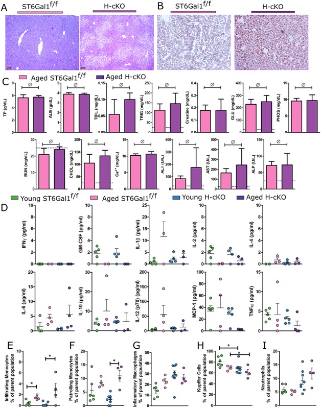
Fig. 8.

Aged H-cKO mice develop spontaneous fatty liver disease. (A) H&E imaging and (B) Oil Red O staining of liver tissues processed from aged mice (approximately 52 weeks of age) demonstrate NAFLD pathology. (C) Blood chemistry analysis revealed no elevation of parameters beyond those expected in a healthy mouse. (D) A 10-plex panel of proinflammatory cytokines was probed for signs of inflammation in the plasma of the ST6Gal1f/f and H-cKO mice at young or old age. (E) Analysis of PMN infiltration into liver revealed no differences. (F–I) Flow cytometric profiling of CD45+CD11b+Ly6C− cells from liver reveals a decrease in Kupffer cells and an increase in proinflammatory associated macrophages in H-cKO mice compared to either their young (8–12 weeks of age) or ST6Gal1f/f controls. Student’s T test, *P < 0.05, ∅: P ≥ 0.05. On each graph, each dot represents one mouse.
