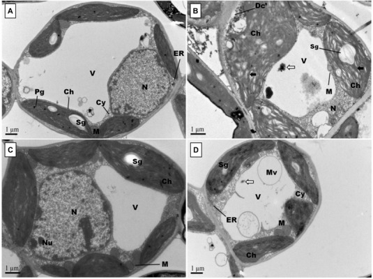
FIGURE 4.

Effects of Pb and GSH on ultrastructure of upland cotton terminal leaves. Transmission electron micrographs represent leaf cell morphology of control (A), 500 μM Pb (B), 50 μM GSH (C) and 500 μM Pb + 50 μM GSH (D) in cotton seedlings. In control cells (A), cytoplasm (Cy) contained large central vacuole (V), nucleus (N) and well developed chloroplasts (Ch) with starch grains (Sg) and plastoglobuli (Pg). Rounded mitochondria (M) and endoplasmic reticulum (ER) can also be detected. After Pb exposure (B), chloroplasts were highly dilated and granal stacks, represented by black arrow (→), were broken. Similarly, electron dense particles and cell debris, represented by white arrow (⟸), was accumulated in the vacuole. GSH-treated leaf cells (C) possessed large chloroplasts, nucleus, nucleolus (Nu), and number of mitochondria were located in between chloroplasts. In GSH + Pb group (D), chloroplasts were dilated but still intact. Moreover, large vacuole with multivesicular structure (Mv), endoplasmic reticulum and nucleus with nucleoli (Nu) were present also.
