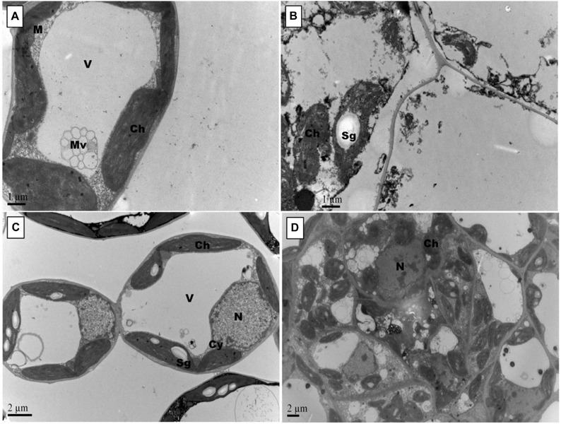
FIGURE 5.

Effects of Pb and GSH on ultrastructure of upland cotton median leaves Transmission electron micrographs represent leaf cell morphology of control (A), 500 μM Pb (B), 50 μM GSH (C) and 500 μM Pb + 50 μM GSH (D) in cotton seedlings. Control cells (A), contained large central vacuole (V), well developed chloroplasts and rounded mitochondria (M). After Pb exposure (B), chloroplasts were highly dilated and plasma membrane detached from cell walls. GSH-treated leaf cells (C) demonstrated large chloroplasts and clearly located nucleus. In GSH + Pb group (D), chloroplasts were dilated but increased in number.
