Figure 1.
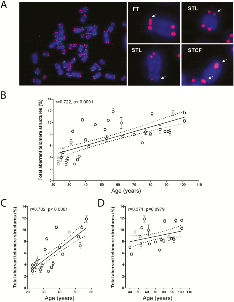
The abundance of aberrant telomeric structures in PBMCs increases with donor age. (A) PBMC metaphase chromosomes (blue) were processed by FISH using a peptide nucleic acid (PNA) complementary to telomeric repeats (red). A representative metaphase spread from a 31-year-old female subject as well as individual metaphase chromosomes displaying aberrant telomeric structures (right) are shown. White arrows point to aberrant telomeric structures. (B) Total aberrant telomere structures (percentage of the observed telomeres) in the PBMCs of each subject. (C) Total aberrant telomere structures (percentage of the observed telomeres) in the PBMCs of each subject belonging to the younger group. (D) Total aberrant telomere structures (percentage of the observed telomeres) in the PBMCs of each subject belonging to the older group. Data are expressed as mean (± SD) of counts from two experiments. Linear regression (continuous line) and 95% confidence band (dotted lines) are shown in B–D. FISH = fluorescence in situ hybridization; FT: fragile telomeres; PBMC = peripheral blood mononuclear cell; STCF = sister telomere chromatid fusions; STL = sister telomeres loss; r = Pearson correlation coefficient; p = statistical significance of Pearson correlation coefficient.

