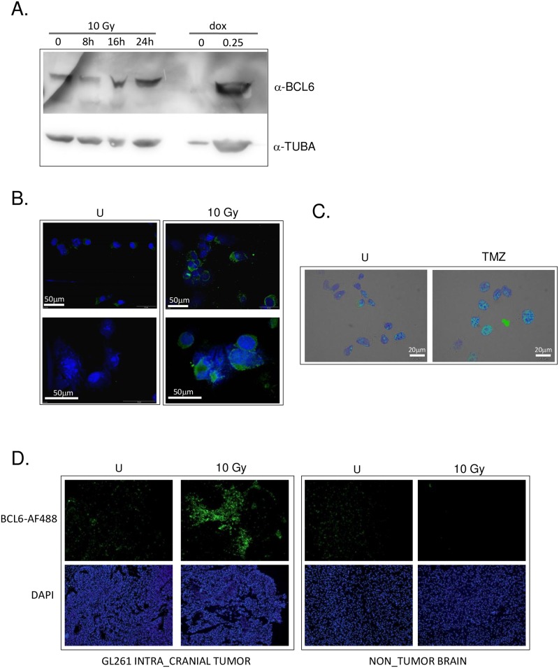
Fig 7. BCL6 was up-regulated by DNA damaging therapy in the transplantable murine glioma model GL261.
A. GL261 cells were treated with 10 Gy of ionizing radiation or 0.25 μM doxorubicin, and harvested at the time point shown (10 Gy) or at 24 hours post-treatment (dox). After electrophoresis and transfer, membranes were cut and blotted with anti-BCL6 (top) or anti-alpha tubulin (bottom). Image representative of at least 3 independent experiments. B. GL261 cells grown on coverslips were treated with 10 Gy of ionizing radiation, then fixed and stained with DAPI (blue) and anti-BCL6 antibody (green). Immunofluorescence images were adjusted only for brightness and contrast, and were adjusted evenly over the entire image. All images were adjusted exactly the same way, and all images representative of at least 3 independent experiments. C. GL261 cells on coverslips were treated with 7 doses of 10 μM TMZ on alternate days, then fixed and stained with DAPI (blue) and anti-BCL6 antibody (green). Immunofluorescence images were adjusted only for brightness and contrast, and were adjusted evenly over the entire image. All images were adjusted exactly the same way, and all images representative of at least 3 independent experiments D. Brains from mice bearing intra-cranial GL261 tumors prior to (U), and 48 hours post irradiation (10 Gy) were collected and BCL6 expression in the tumor and the normal brain determined by immunofluorescence microscopy using Alexa-488-labelled anti-BCL6 antibody. Nuclei were imaged with DAPI. All data shown are representative of at least 3 independent experiments. Cropped gel images retain all bands, immunofluorescence images were adjusted only for brightness and contrast, and were adjusted evenly over the entire image.

