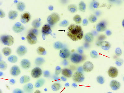
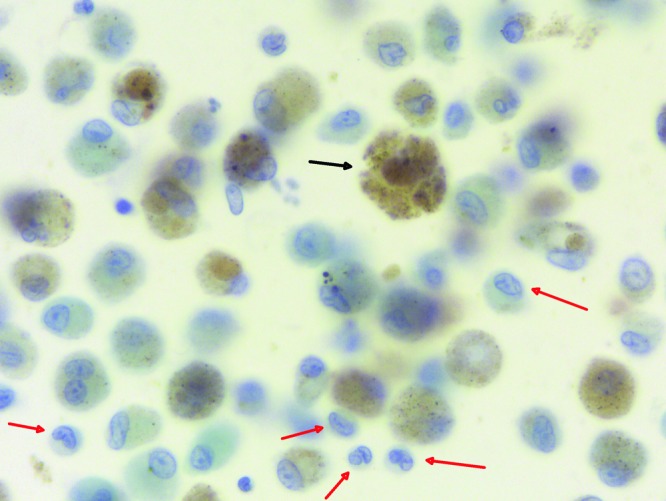
Fig. 16.3.


Alveolar macrophages (black arrows) with smoker’s inclusions and Langerhans’ cells in pulmonary Langerhans’ cell histiocytosis (red arrows). Note the notches of the nuclei of Langerhans’ cells (Courtesy of Dr. Henry Budihardjo Welim, Institute of Pathology Hemer)
