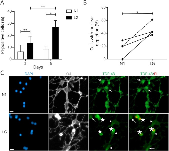
Figure 5. Nuclear depletion of TDP-43 in human primary oligodendrocytes cultured in vitro under LG conditions.
Data were derived from 3 adult cases and 1 pediatric case with no history of MS. (A) Cell death of human oligodendrocytes was assessed using PI staining at 2 and 6 days under optimal (N1) and metabolic stress (LG) culture conditions. (B) Depletion of nuclear expression of TDP-43 in cultures of oligodendrocytes after 2 days of treatment with LG compared with N1 condition. Solid lines (adult cases) and dashed line (pediatric case) connect cultures of oligodendrocytes obtained from the same biopsy tissue. (C) Representative images showing immunostained oligodendrocytes 2 days under N1 and LG conditions. From left side: DAPI (blue), O4 (gray), TDP-43 (green), and TDP-43 merged with PI (green and red, respectively). Arrows show cells with nuclear expression of TDP-43; arrowheads show TDP-43 nuclear-depleted PI− cells; stars show TDP-43 nuclear-depleted PI+ cells. Scale bar = 10 μm. *p < 0.05, **p < 0.01. DAPI = 4′,6-diamidino-2-phenylindole; LG = low glucose; PI = propidium iodide; TDP-43 = transactivation response DNA-binding protein of 43 kDa.

