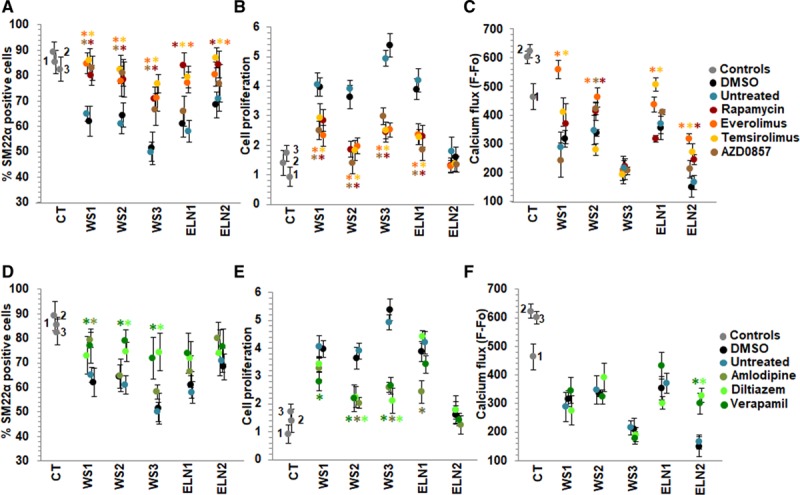
Figure 5.

Effect of candidate drugs on smooth muscle cell (SMC) differentiation, proliferation, and calcium flux. A–C, mTOR (mammalian target of rapamycin) inhibitors. A, The percentage of SM22α (smooth muscle 22α) positive cells measured by high content imaging showed that dimethyl sulfoxide (DMSO)-treated elastin insufficiency (EI) patient SMCs express 50%–70% SM22α (black dots) similar to untreated patient cells (blue) in contrast to 80%–90% expressed in control SMCs (gray). Rapamycin (dark red), everolimus (orange), and temsirolimus (yellow) increased % of SM22α positive cells in all patients when compared to DMSO treatment (black). AZD0857 (brown) only increased SMC differentiation in Williams syndrome (WS) patient SMCs. B, All 4 mTOR inhibitors decreased SMC proliferation in all WS and in ELN (elastin)-1 cells. ELN2 cells were not hyperproliferative and did not show any further change in proliferation with mTOR inhibitors. C, Endothelin-induced calcium flux was increased by everolimus in WS1, WS2, ELN1, and ELN2-SMCs compared with DMSO-treated cells. Rapamycin only improved calcium flux in two patients (WS2, ELN2), temsirolimus in 3 patients (WS1, ELN1, ELN2), and AZD0857 in 1 patient (WS2). WS3 did not respond to any mTOR inhibitor. D–F, Calcium channel blockers. D, Verapamil (dark green) and diltiazem (bright green) increased % of SM22α positive cells only in 3 and 2 WS patients, respectively, but not in elastin mutation patients. E, Verapamil and diltiazem decreased SMC proliferation only in 3 and 2 WS patients, respectively, not in elastin mutation patients. Amlodipine (military green) treatment was associated with cell death (data not shown). F, Verapamil and diltiazem improved endothelin-induced calcium flux only in ELN2 patient SMCs (n=3 independent experiments, using 3 technical replicates for each experiment). A–F, Supporting data are shown in Table IIIB in the Data Supplement. *P<0.05, drug treatment vs DMSO.
