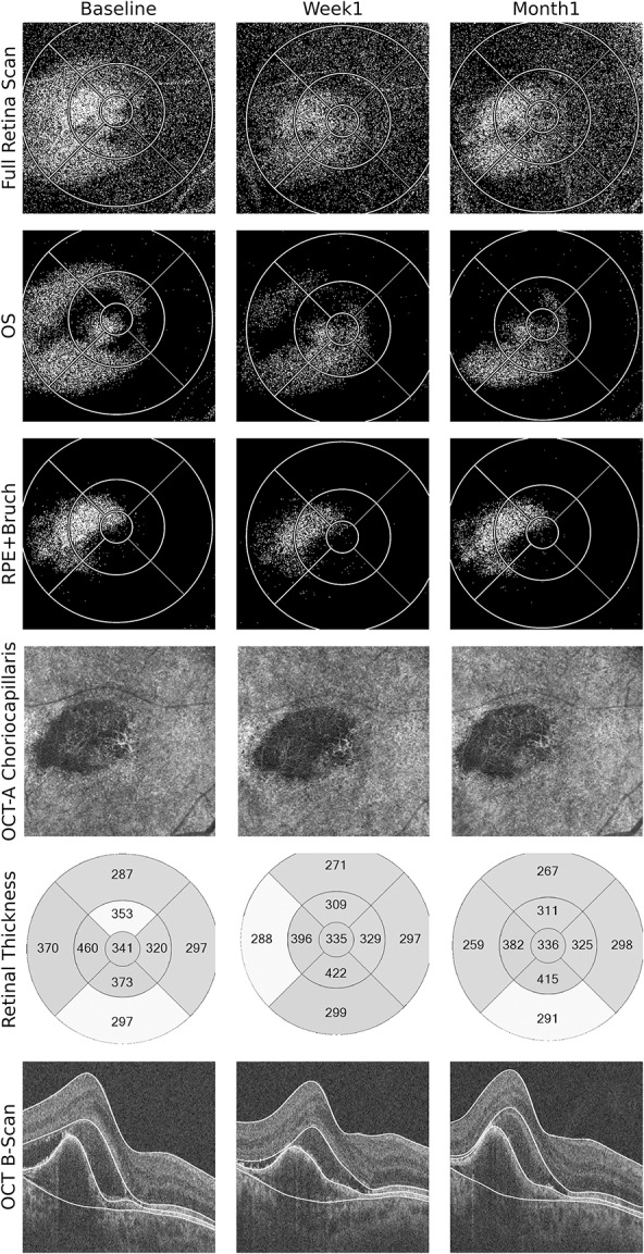
Fig. 2.

Multimodal OCT-based imaging of the right eye of a patient with exudative age-related macular degeneration immediately before, 1 week, and 1 month after IVA injection. Optical coherence tomography leakage map of the full retina scan in the 1st row shows the extension of fluid accumulation and its change during follow-up. This change took place mainly in the OS and RPE–Bruch layers (2nd and 3rd rows). One can see a steady decrease in fluid distribution in the OS layer; however, in RPE–Bruch layer, there seems to be more fluid in 1st month compared with the 1st week. Accordingly, the conventional B-scans show a decrease in subretinal fluid, but the fibrovascular PED seems to increase again after 1 month (6th row). The OCTA analysis also shows a decrease in vascularization of the CNV after 1 week; however, there is repermeabilization of the vascular network in the Month 1, with a flow signal similar to baseline (4th row). Association of noninvasive OCTA and OCT-L thus show structural changes occurring before significant RT change and provide insight into a case that might need early retreatment to control the exudative membrane. Optical coherence tomography leakage segmentation used in the layers depicted is marked in white in the SD-OCT images. Optical coherence tomography angiography segmentation was customized for better visualization of the neovascular network.
