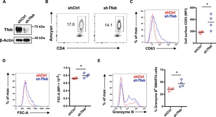
Fig. 6. Tfeb deficiency reproduces human aged phenotypes in a mouse model in vivo.

Tfeb shRNA retrovirally transduced Amcyan+ LCMV-specific naïve SMARTA transgenic CD4+ T cells (2 × 10⁵) were adoptively transferred into CD45.2+ naïve recipients. Mice were infected with 2 × 10⁵ plaque-forming units of LCMV Armstrong. On day 5 after infection, spleens were harvested and analyzed. Fluorescence-activated cell sorting plots are gated on CD4+ Amcyan+ SMARTA cells. (A) Tfeb silencing in sorted CD4+ Amcyan+ SMARTA cells was confirmed by Western blotting. (B) Contour plot showing percentage of retrovirally transduced Amcyan+ SMARTA cells on day 5 after LCMV infection. (C) Representative histogram of CD63 cell surface expression on transduced SMARTA cells on day 5 after infection (left) and cumulative data from four mice in each group (right). (D) Representative histogram of FSC-A (left) and cumulative data from four mice in each group (right). (E) Representative histogram of intracellular granzyme B (left) and cumulative data of percentage of granzyme B+ cells in Amcyan+ SMARTA cells (right) from four mice in each group. Data are representative of one of two independent experiments each with n = 4 mice. *P < 0.05.
