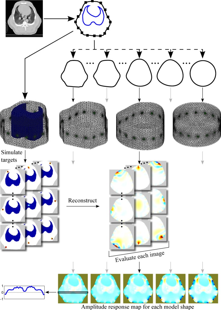
Fig. 1.
Overview of the methods. A CT slice in the electrode plane is segmented to obtain the boundary shape, the contour of the lungs and the electrode positions. An FEM with a lung contrast conforming to the CT slice is created and used to simulate a number of targets covering the whole body. Several homogeneous models with distorted shape are created and used for reconstruction. Based on the individual target reconstructions, a map representing performance metric as a function of position is constructed for each model shape.

