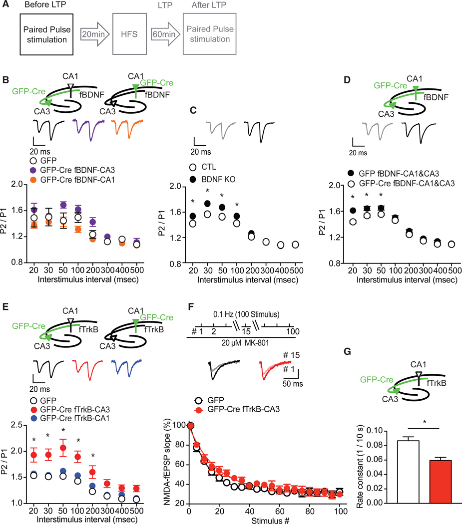
Figure 4. Deletion of Presynaptic TrkB Alters Release Probability.
(A) Experimental protocol.
(B) Compared to GFP controls, paired-pulse ratio (PPR) (P2/P1) before LTP was not altered by deletion of BDNF in either CA1 or CA3 (two-way ANOVA: F(2,192) = 12.21, p = 0.3474 for group; F(7,192) = 22.1, p < 0.0001 for interstimulus interval).
(C) PPR was increased in hippocampal slices (n = 7) of Bdnf knockout (KO) mice, compared to littermate CTLs (n = 7) (two-way ANOVA: F(1,88) = 24.78, p < 0.0001 for group; F(7,208) = 12.81, p < 0.0001 for interstimulus interval; Sidak’s post hoc tests: CTL versus KO, 20 ms, p = 0.0388; 30 ms, p = 0.0006; 50 s, p = 0.0054; 100 ms, p = 0.0491).
(D) Deletion of BDNF in both CA1 and CA3 regions increased PPR (two-way ANOVA: F(1,184) = 40.97, p < 0.0001 for group; F(7,184) = 22.23, p < 0.0001 for interstimulus interval: Sidak’s post hoc tests: GFP versus GFP-Cre, 20 ms, p < 0.0001; 30 ms, p = 0.0046; 50 ms, p = 0.0194) before LTP. Representative traces show pulse 1 following by pulse 2 with 20-ms interstimulus interval.
(E) Compared to GFP controls, deletion of TrkB in CA1 had no impact on PPR before LTP; however, selectively deleting TrkB from CA3 enhanced PPR (two-wayANOVA: F(2,208) = 57.4, p < 0.0001 for group; F(7,208) = 35.64, p < 0.0001 for interstimulus interval; Sidak’s post hoc tests: CTL versus GFP-Cre Ntrk2fl/fl -CA3, 20 ms, p = 0.0003; 30 ms, p = 0.0001; 50 ms, p < 0.0001; 100 ms, p < 0.0001; 200 s, p = 0.0008) before LTP. Representative traces show pulse 1 following by pulse 2 with 20-ms interstimulus interval.
(F) Top, To measure release probability, NMDA-fEPSP was recorded from the CA3-CA1 Schaffer collateral pathway, using 0.1-Hz stimulation, before and afterMK-801 bath application. Traces show responses of the 1st and the 15th stimulus. NMDA-fEPSP amplitudes evoked every 10 s in the presence of MK-801 decayed slower in slices with deletion of TrkB in CA3 (n = 7) compared to GFP (n = 8).
(G) Rate constant of NMDA-fEPSP amplitudes in the presence of MK-801 was significantly lower in slices with deletion of TrkB in CA3 than GFP (t(13) = 3.631, p = 0.003).
*p < 0.05. Data are mean ± SEM.

