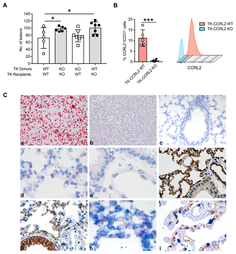
Figure 4. CCRL2 expressed by non-hematopoietic cells plays a role in the control of tumor growth.
(A) TK-CCRL2 WT and TK-CCRL2 KO mice were lethally irradiated, then reconstituted with total bone marrow cells from TK-CCRL2 WT and TK-CCRL2 KO donor mice. Eight weeks after reconstitution, tumor development was induced by intranasal inoculation with 2.5x10⁷ infectious particles of Ad5CMVCre recombinase, then mice were sacrificed after 10 weeks. The number of lesions is indicated. *p<0.05, by Generalized Linear Mixed Model in family Poisson (TK-CCRL2 WT recipients of TK-CCRL2 WT donors, n=5; TK-CCRL2 KO recipients of TK-CCRL2 KO donors, n=6; TK-CCRL2 WT recipients of TK-CCRL2 KO, n=7; TK-CCRL2 KO recipients of TK-CCRL2 WT donors, n=7) (B) CCRL2 expression on CD31+ in CD45- gated cells of lungs from tumor-bearing mice and representative histograms. ***p<0.001, TK-CCRL2 WT (n=5) vs. TK-CCRL2 KO (n=4) by Student t-test. (C) CCRL2 mRNA expression was investigated by RNAscope. The CCRL2 mRNA probe was tested on formalin-fixed blocks from CCRL2 transfected L1.2 cells as a positive control (a) and L1.2 mock cells (b) and CCRL2 KO lung as negative controls (c). CCRL2 mRNA in alveolar wall (d), endothelial cells of large vessel (e) and immunohistochemistry for CD31 (f) and cytokeratin (g) staining of CCRL2 WT lung. (h) CCRL2 mRNA co-stained with anti-cytokeratin antibody (i) double immunohistochemistry of human lung adenocarcinoma (LUAD) biopsy with anti-CCRL2 and anti-ERG moAbs shows double positive endothelial cells in the peritumoral area.

