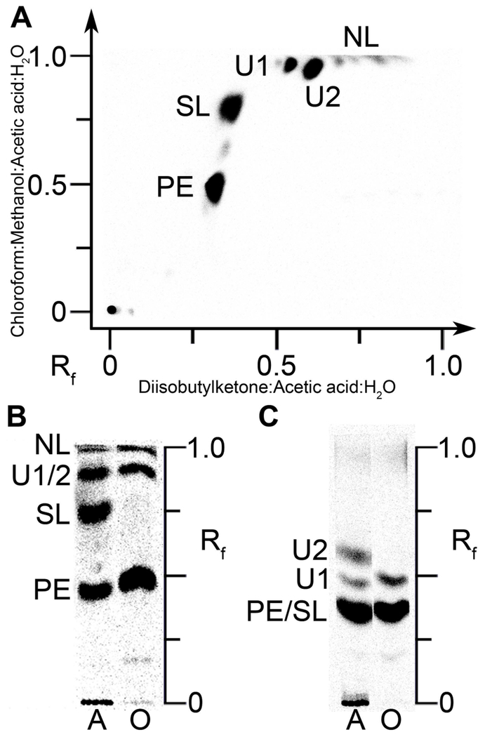
Figure 1. Polar lipids of A. finegoldii.
PE, phosphatidylethanolamine; SL, sulfonolipid; U1 and U2 were not identified; NL, neutral lipids. PE and SL were identified by mass spectrometry analysis as shown in subsequent figures.
A. A. finegoldii was labeled with [14C]acetate and the lipid extract was separated by two-dimensional chromatography on Silica Gel H thin layers using chloroform:methanol:acetic acid:water (80/25/10/2, v/v) in the first dimension followed by diisobutylketone:acetic acid:water (80/55/15, v/v) in the second dimension. The labeled lipids were visualized with a PhosphorImager system.
B. Lipid extracts from cells labeled with either [14C]acetate (A) or [14C]oleate (O) were separated using the first-dimension solvent system.
C. Separation of [14C]acetate (A) or [14C]oleate (O) labeled lipids using the second-dimension solvent system.

