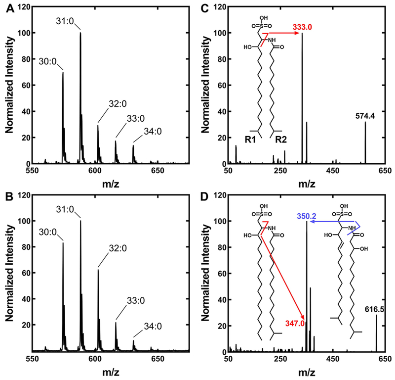
Figure 4. Molecular species of A. finegoldii SL.
The total apparent number of carbons in the SL that are derived from fatty acids are indicated.
A. A representative mass spectrum of the SL fraction of A. finegoldii grown in DSM Medium 104.
B. A representative mass spectrum of SL from A. finegoldii grown in WCAB minimal medium.
C. A representative MS/MS spectrum of the 30:0 SL molecular species. Inset, the structure of the single 30:0 SL species present that was derived from two 15:0 fatty acids (sulfobacin B). The diagnostic peak is indicated along with the fragmentation pattern. R1 of SL is the fatty acid condensed with cysteic acid, and R2 is the fatty acid attached to the amine.
D. A representative MS/MS spectrum of the 33:0 SL molecular species. Inset, a representative MS/MS spectrum showing the structures of the two SL molecules that comprise the 33:0 SL molecular species. The minor species is SL derived from 16:0 and 17:0 fatty acids. The major species is derived from a trans-2–15:0 and a 3-hydroxy-17:0 fatty acids (flavocristamide A). The 32:0 SL molecular species is also a mixture of two SL species (not shown).

