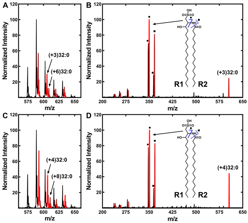
Figure 6. Incorporation of extracellular myristate and palmitate into SL.
A. finegoldii was labeled with either [d3]14:0 or [d4]16:0, and the lipids were extracted as described under “Experimental Procedures.” Mass spectra of the SL were obtained in both cases and fragmented to determine the positional distribution of the incorporated fatty acids into the SL molecule. R1 of SL is the fatty acid condensed with cysteic acid, and R2 is the fatty acid attached to the amine.
A. SL from cells labeled with [d3]14:0.
B. Fragmentation of the (+3)32:0-SL molecular species. Inset, ● and ■ indicate the two diagnostic fragments arising from (+3)32:0-SL as shown in the structure diagram.
C. SL molecular species from cells labeled with [d4]16:0.
D. Fragmentation of the (+4)32:0-SL molecular species. Inset, ● and ■ indicate the two diagnostic fragments arising from (+4)32:0-SL as shown in the structure diagram. SL molecular species (A, C) or fragments (B, D) containing a deuterium label are indicated in red.

