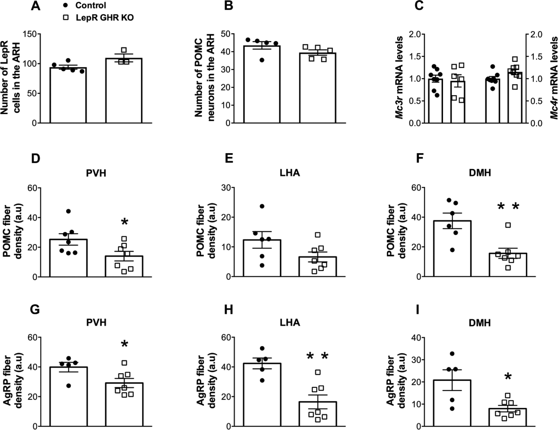
Fig. 5. GHR ablation in LepR-expressing neurons affects the neuronal projections of the arcuate nucleus.

A. Number of LepR-expressing cells in the ARH of control (n = 5) and LepR GHR KO (n = 3) mice. B. Number of POMC neurons in the ARH of control (n = 5) and LepR GHR KO (n = 5) mice. C. Hypothalamic mRNA levels of Mc3r and Mc4r in control (n = 8) and LepR GHR KO (n = 6–8) mice. D-F. Quantification of POMC fiber density in the PVH (A), LHA (B) and DMH (C) of control (n = 6–7) and LepR GHR KO (n = 7) mice. G-I. Quantification of AgRP fiber density in the PVH (D), LHA (E) and DMH (F) of control (n = 5) and LepR GHR KO (n = 7) mice. Mean ± s.e.m. Two-tailed Student’s t-test. * P < 0.05, ** P < 0.01 vs. LepR GHR KO.
