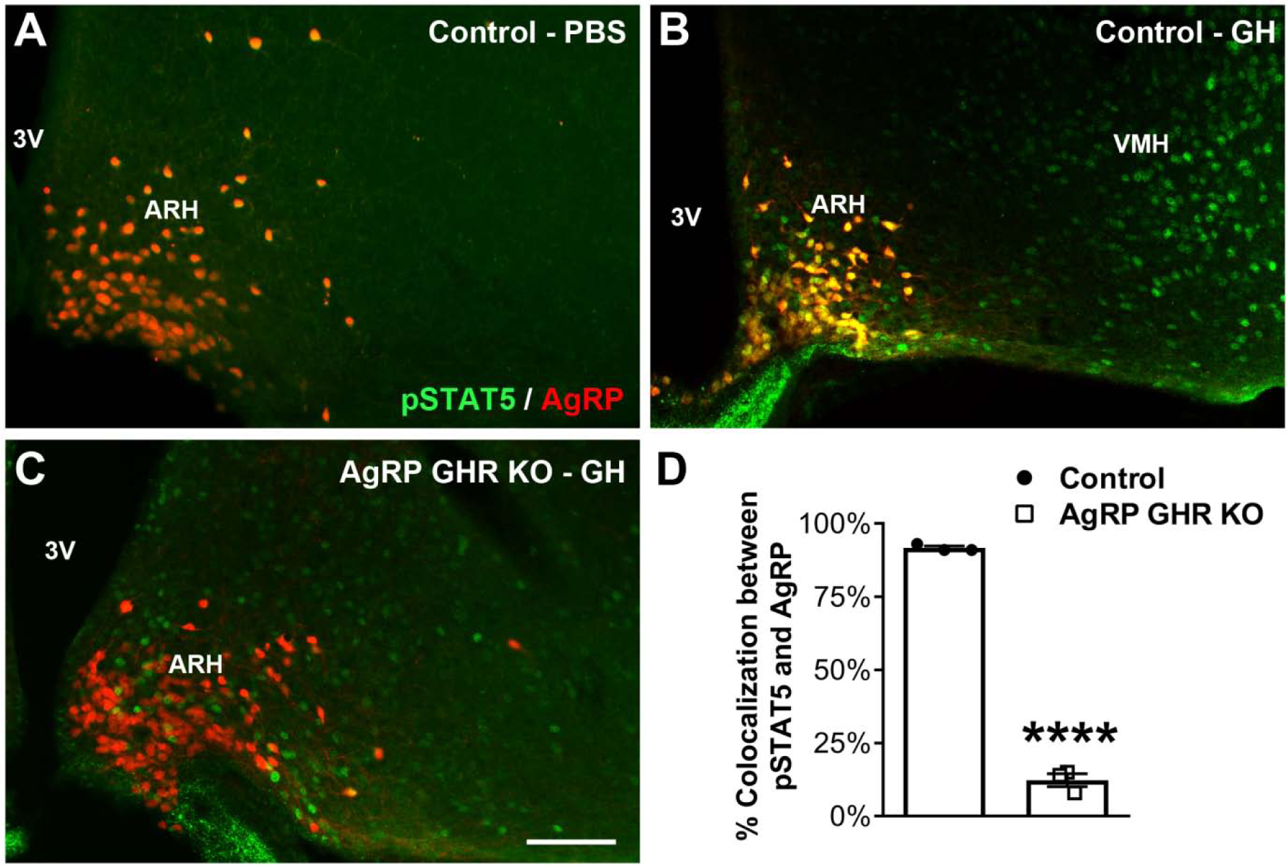
Fig. 7. AgRP-expressing neurons are responsive to GH.

A-C. Epifluorescence photomicrographs showing double-labeling immunofluorescence staining of pSTAT5 (green) and AgRP (red tdTomato protein) in the arcuate nucleus of the hypothalamus (ARH) of PBS-injected control mice (A), GH-injected control mice (B) and GH-injected AgRP GHR KO mice (C). Yellow represents double-labeled cells. D. Percentage of colocalization between pSTAT5 and AgRP in the ARH of GH-injected control mice (n = 3) and GH-injected AgRP GHR KO mice (n = 3). Mean ± s.e.m. Abbreviations: 3V, third ventricle; VMH, ventromedial nucleus of the hypothalamus. Scale bar = 100 μm. **** P < 0.0001 vs. AgRP GHR KO.
