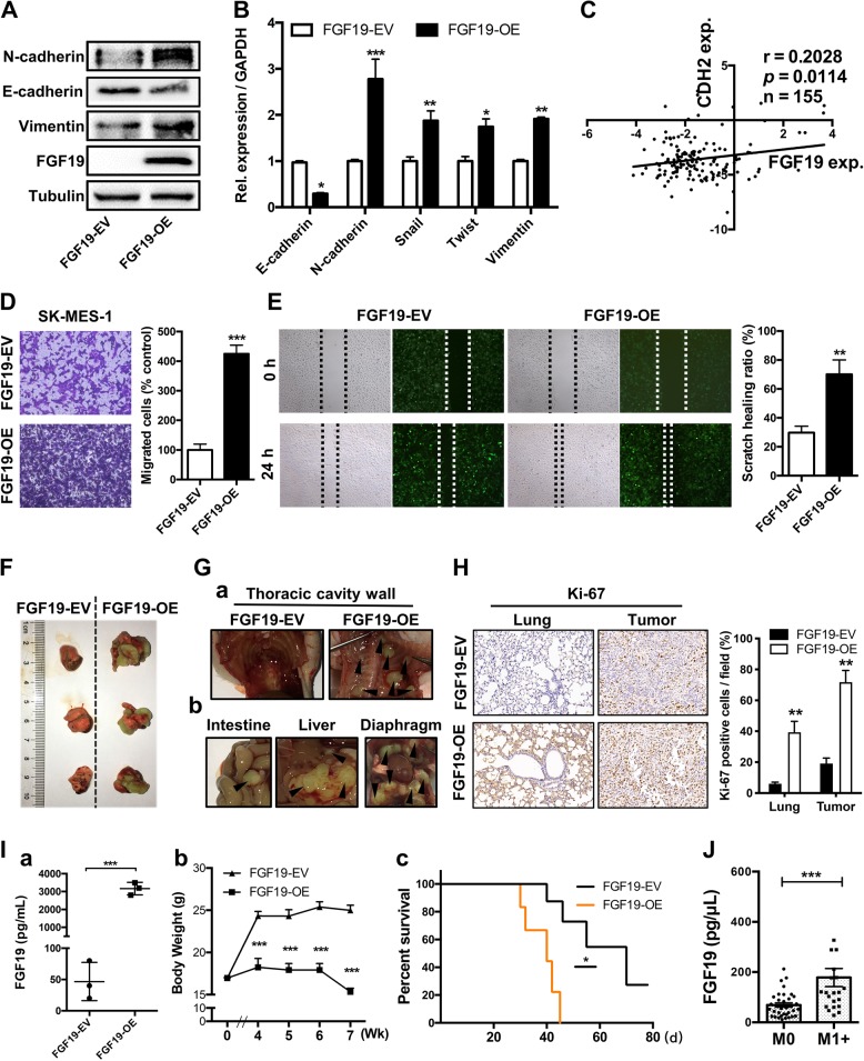
Fig. 4. Overexpression of FGF19 promotes metastasis of LSQ cells in vitro and in vivo.
a Protein and b mRNA expression of EMT markers in the cells treated with FGF19-EV or FGF19-OE for 48 h in SK-MES-1 cells. c Analysis of correlation between CDH2 and FGF19 from Oncomine TCGA datasets. The images of migration abilities in FGF19-OE transfected cells in trans-well (d) and wound healing (e) assays, respectively. f Overexpression of FGF19 increased tumor growth in vivo in orthotopic lung cancer models. Representative image of primary tumors in the left lungs of orthotropic models on day 21 from each group after implantation of SK-MES-1 cells transfected with FGF19-OE and FGF19-EV. g Metastatic tumors in other organs. h Representative images of IHC analysis of Ki-67 expression in lung and tumors. Right panel: Quantification of Ki-67 expression. i (a) Evaluation of FGF19 expression in serum samples from two groups. (b) Mean primary tumor body weight and (c) survival curve for the mice in each treatment group evaluated. j Within the 57 NSCLC patients’ group, the serum levels of FGF19 in patients with metastasis (n = 34) were shown side by side with those of patients without metastasis (n = 23). Data are represented as mean and SEM from three independent experiments. *p < 0.05; **p < 0.01; ***p < 0.001.

