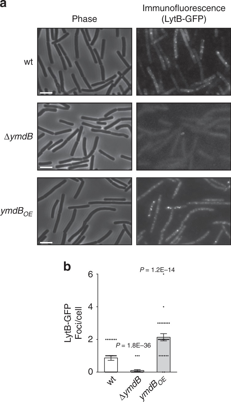
Fig. 2. LyB localizes to the cell surface in YmdB-dependent manner.

a, b Bs wt (AB132: lytB-gfp), ∆ymdB (AB134: lytB-gfp, ΔymdB) and ymdBOE (AB144: lytB-gfp, ΔymdB, amyE::Phyper-spank-ymdB) strains were spotted onto poly l-lysine coated coverslips, treated with anti-GFP primary antibodies and FITC-conjugated secondary antibodies and visualized by fluorescence microscopy. Shown are phase contrast images (left panels) and respective immunofluorescence images (right panels). Scale bars represent 2 μm (a). Graph presents quantification of the average number of LytB-GFP foci displayed per cell. Shown are mean ± SEM and P values (unpaired Student’s t test) of at least three independent experiments (ncells = 600) (b). Source data are provided as a Source Data file.
