Figure 1.
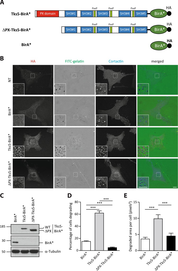
Tks5-BirA* is active and localizes in invadopodia. (A) Schematic representation of BirA* fusion proteins used in this study. The PX, SH3 and Proline rich (PxxP) domains of Tks5 are depicted. All fusion proteins have a HA tag at their C-terminus. (B) Representative images of MDA-MB-231 cells stably expressing wild-type Tks5 or Tks5 lacking its PX domain (ΔPX) fused to the biotin ligase BirA* or BirA* alone, and of non-transfected (NT) cells, seeded on fluorescently-labeled gelatin (FITC-gelatin) for 4 hours. Cells were fixed and stained with anti-HA and -cortactin antibodies. Invadopodia were identified thanks to cortactin labeling. Active invadopodia were identified thanks to degradation area (dark spots) in FITC-gelatin. The white-boxed regions are shown enlarged in the bottom insets. Scale bars represent 5μm. (C) Expression levels of BirA* fusion proteins in MDA-MB-231-derived cells described in (B) was assessed by western blotting using an anti-HA antibody. α-Tubulin was used as a loading control. (D,E) Tks5-BirA* fusion proteins are functional. The ability of MDA-MB-231 cells expressing Tks5-BirA* fusion proteins, described in (B), to degrade fluorescently-labeled gelatin was analyzed. The percentage of degrading cells (D) and the degraded area per cell (E) are represented as the mean ± SEM of three independent experiments. ***p ≤ 0.001. Full-length blots are presented in Supplementary Fig. 2.

