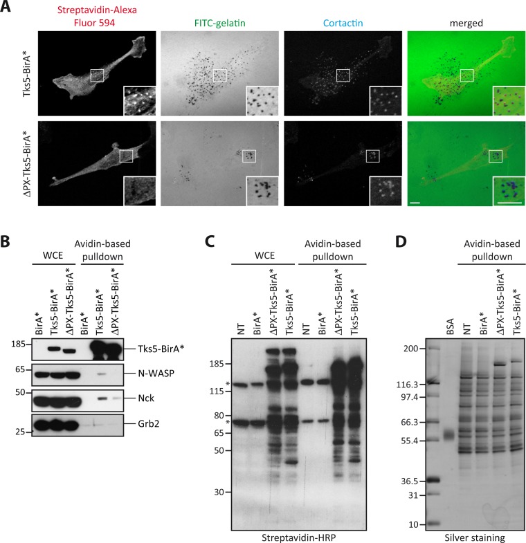
Figure 2.
Selective biotinylation of invadopodia proteins by Tks5-BirA*. (A) Representative images of MDA-MB-231 cells stably expressing Tks5-BirA* or ΔPX-Tks5-BirA* seeded on fluorescently-labeled gelatin (FITC-gelatin) in the presence of biotin for 16 hours. Cells were fixed and stained for cortactin as a marker of invadopodia and streptavidin coupled to Alexa Fluor 594 to reveal biotinylated proteins. The white-boxed regions are shown enlarged in the bottom insets. Scale bars represent 5μm. (B) Analysis of the biotinylation of known Tks5 interactors and of well characterized invadosome components. MDA-MB-231 cells stably expressing BirA*, Tks5-BirA* or ΔPX-Tks5-BirA* were seeded on gelatin in presence of biotin for 16 h. After cell lysis, biotinylated proteins were isolated by affinity capture using avidin-coated beads. The presence of proteins of interest was assessed in whole cell extract (WCE) or after avidin-based pulldown by western blotting with specific antibodies. (C) Isolation of proteins biotinylated by Tks5-BirA* and ΔPX-Tks5-BirA*. MDA-MB-231 cells stably expressing Tks5-BirA*, ΔPX-Tks5-BirA* or BirA*, and non-transfected (NT) cells were seeded on gelatin and treated with biotin for 16 h before cell lysis. Biotinylated proteins were pulled down via avidin-coated beads. The presence of biotinylated proteins was assessed by western blotting in whole cell extract (WCE) or after avidin-based pulldown (one-twentieth of the total pulldown was used) using HRP-coupled streptavidin. (*) corresponds to intrinsically biotinylated proteins. (D) One-tenth of the samples shown in (C) was resolved by SDS-PAGE for silver staining of the isolated proteins; the rest was analyzed by mass spectrometry. Full-length blots/gels are presented in Supplementary Fig. 2.

