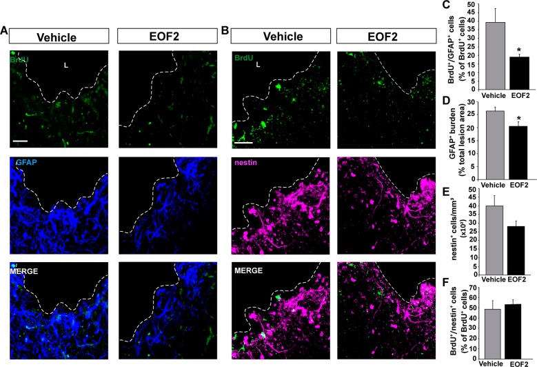
Fig. 6. Local administration of EOF2 reduce glial differentiation in the injured cortex.
Representative confocal microphotographs of the area surrounding cortical lesion in mice brain processed for the immunohistochemical detection of the glial marker GFAP (a) and for the undifferentiated progenitors marker nestin (b). Mechanical cortical lesions were unilaterally performed in the primary motor cortex of adult mice, and osmotic minipumps were implanted to locally deliver vehicle or EOF2 (5 µM) for 14 days. All mice were intraperitoneally-injected with BrdU the last day of treatment as described in methods. Scale bar represent 50 µm. The dotted line indicates the limit of the lesion (L). c Graph shows the percentage of BrdU+ cells that co-express the glial marker GFAP in the peri-lesional area of the indicated animal groups. Data shown are the mean ± S.E.M.; n = 6 animals per group. Statistical analysis: *p = 0.0492 in two tailed unpaired Student’s t test comparing EOF2 with the control. d Graph shows the GFAP burden, representing the glial scar, expressed as a percentage of the total lesion area. Data shown are the mean ± S.E.M.; n = 6 animals per group. Statistical analysis: *p = 0.0310 in two tailed unpaired Student’s t test comparing EOF2 with the control. e Quantification of nestin+ cells per mm3 in the peri-lesional area of the indicated animal groups. Data shown are the mean ± S.E.M.; n = 6 animals per group. f Graph shows the percentage of BrdU+ cells that co-express the neural precursor marker nestin in the peri-lesional area of the indicated animal groups. Data shown are the mean ± S.E.M.; n = 6 animals per group.

