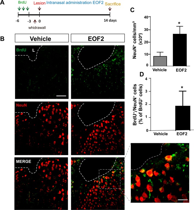
Fig. 8. Intranasal administration of EOF2 induces neuronal differentiation in the peri-lesional area after brain injury.
a Scheme representing the experimental procedure followed to label proliferating neural precursors with BrdU exclusively in neurogenic niches and not in the injured area: mice received BrdU injections on days 6, 5, and 4 before performing the cortical injury; then we waited three more days to allow for complete withdrawal of BrdU from the animal organism. Lesion was performed on day 0. Intranasal administration of EOF2 (5 µM) or only vehicle was performed for 14 days. b Representative confocal microscopy images of the injured cortex of adult mice after bearing cortical lesions and the intranasal administration of EOF2 or only vehicle. The dotted line indicates the limit of the lesion (L) and the scale bar represent 100 µm in the low magnification pictures and 20 µm in the high magnification pictures. c Graph shows the number of neuronal cells marked with NeuN per mm3 in the peri-lesional area of the indicated animal groups. Data shown are the mean ± S.E.M.; n = 6 animals per group. Statistical analysis: *p = 0.0339 in two tailed unpaired Student’s t test comparing EOF2 with the control. d Percentage of BrdU+ cells that co-expressed the neuronal marker NeuN in the peri-lesional area. Data shown are the mean ± S.E.M.; n = 6 animals per group. Statistical analysis: *p = 0.0411 in two tailed unpaired Student’s t test comparing EOF2 with the control.

