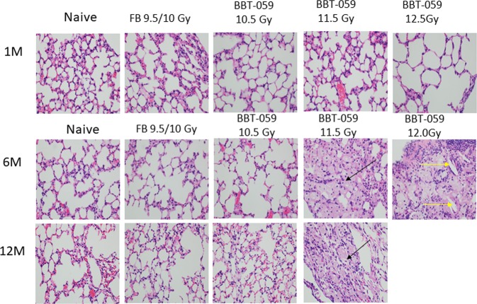
Figure 5.
Lung histology demonstrates cholesterol clefts and Histiocytes. Differences between the histiocytic pneumonitis and the normal lungs. These photos of lungs taken at 40× magnification at the objective (total 400×) Photos BBT-059 11.5–6 M, BBT-059 11.5–12 M; and BBT-059 12–6 M; all show histiocytic pneumonitis. The large “foamy” cells identified with arrows are all histiocytes. The yellow arrow identifies a “cholesterol cleft”. They commonly occur in areas of the lungs with these foamy macrophages, often in association with a lot of proteinaceous material. Please note, it is possible that the histiocytic pneumonitis is simply a background lesion that would have occurred in these animals regardless of exposure.

