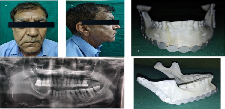
Fig. 3.
a, b Preoperative frontal view and right facial profile showing asymmetry and swelling of face in the left posterior region of mandible; c OPG showing multilocular radiolucent lesion extending from the distal aspect of right first premolar up to the sigmoid notch involving the condyle, coronoid, anterior, posterior border of ramus and lower border of mandible; d, e 3D model obtained to facilitate plate adaptation prior to resection

