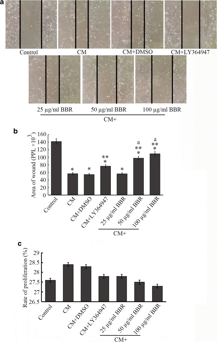
Fig. 3.
Cultured HCoEpiC cells are wounded by a 200-μl pipette tip and observed by phase contrast microscopy (100 ×) after different treatments for 24 h (a). To measure the area of the wound, software (ImageJ) was used, and the area of wound was presented as pixels per inch (PPI), *P < 0.05 versus control, **P < 0.05 versus CM group, and aP < 0.05 versus CM + 25 μg/ml BBR group (b). The error bar represents the SD (n = 3). In addition, we conducted a CCK-8 assay to observe the effect of CM or BBR on cell proliferation. As shown in c, the percentage of proliferation of the CM or DMSO group was mildly higher than that of the control group, and there was no significant difference between the two groups. Similarly, a significant difference between the BBR groups and the CM or control group was not observed, suggesting that the effect of CM or BBR treatment on proliferation was not significant

