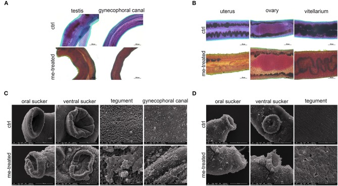
Figure 2.
Morphological changes in male and female of S. japonicum in vitro. Adult worm pairs were incubated with myricetin (me) at 600 μM for 24 h, and female and male worms were separated for analysis. Adult male (A) and female (B) were stained with acetocarmine-fast green and observed under Leica microscope. (C) Scanning electron microscopy (SEM) analysis of the oral sucker, ventral sucker, tegument and the special structure of male worms, gynecophoral canal of S. japonicum following treatment with myricetin. Scale bars: (A) upper left row, 100 μm; lower right row, 200 μm (B) 50 μm and (C) The oral sucker and ventral sucker of male worms, 100 μm; (D) The oral sucker and ventral sucker of female worms, 40 μm; The tegument and gynecophoral canal, 20 μm. ctrl, adult worms were incubated in RPMI 1,640 medium with 1% DMSO; me-treated, adult worms treated with myricetin.

