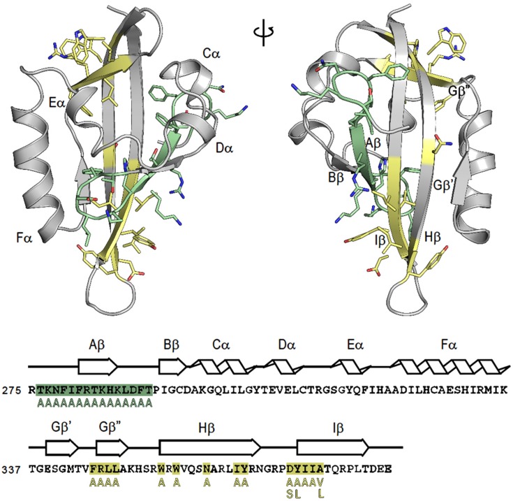
Figure 1.
Structure and sequence of the mouse aryl hydrocarbon receptor (AhR) (Pubmed sequence: NP_038492.1) and map of generated mutations. The structure of the AhR PASB is taken from the homology model [33]. Structural elements of the AhR PASB homology model are indicated both in the structure and above the sequence. Mutants in A and B strands (N-terminal) are colored in green, while mutants in the G, H, and I strands (C-terminal) are colored in yellow.

