Figure 1.
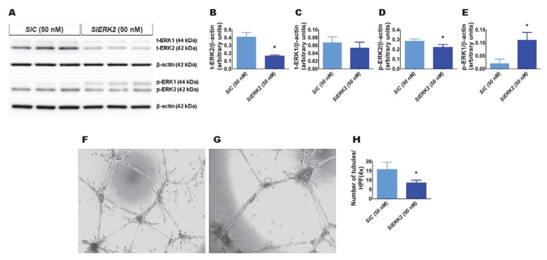
The effects of ERK2 knockdown on human pulmonary microvascular endothelial cell (HPMEC) tubule formation. Control- or ERK2-siRNA-transfected HPMECs were harvested for protein expression and tubule formation assays. (A) The determination of total (t)- and phosphorylated (p)-extracellular signal-regulated kinase (ERK)2 and ERK1 protein levels in control (SiC)- and ERK2 (SiERK2)-siRNA-transfected cells by immunoblotting. (B–E) Quantification and normalization of t-ERK2 (B), t-ERK1 (C), p-ERK2 (D), and p-ERK1 (E) band intensities to those of β-actin. (F–H) Representative photographs showing the tubule formation of cells transfected with control (F) and ERK2 (G) siRNAs. (H) Quantification of tubule formation in control (SiC) and ERK2 (SiERK2) siRNA-transfected cells. Values are presented as mean ± SD (n = 3/group for the protein expression studies and n = 7–8/group for the tubule formation assay). Significant differences between control siRNA- and ERK2 siRNA-transfected groups are indicated by *, p < 0.05 (t-test).

