Figure 2.
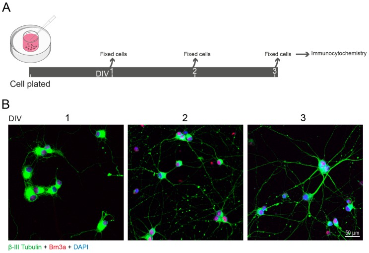
Neurite growth of RGCs in culture. (A) Schematic representation of the experimental design. Retinas were dissected from Wistar rats at PND 5 and nearly pure RGC cultures (~93% purity assessed with anti-RBPMS antibody; Abcam, Cat. # ab194213, 1:500) were obtained by sequential immunopanning, as previously described [8,9]. RGCs were cultured for 1 day in vitro (DIV1), DIV2 and DIV3, followed by fixation in paraformaldehyde and processed for immunocytochemistry. (B) RGCs were identified by immunolabeling for Brn3a (red, Millipore, Cat. # MAB1585, 1:500), a transcription factor expressed only by these cells in the retina. The neurites, labelled with an antibody that recognizes β-tubulin III (green, BioLegend, Cat. # 802001; 1:1000), extended during the period in culture. Nuclei were stained with DAPI (blue).

