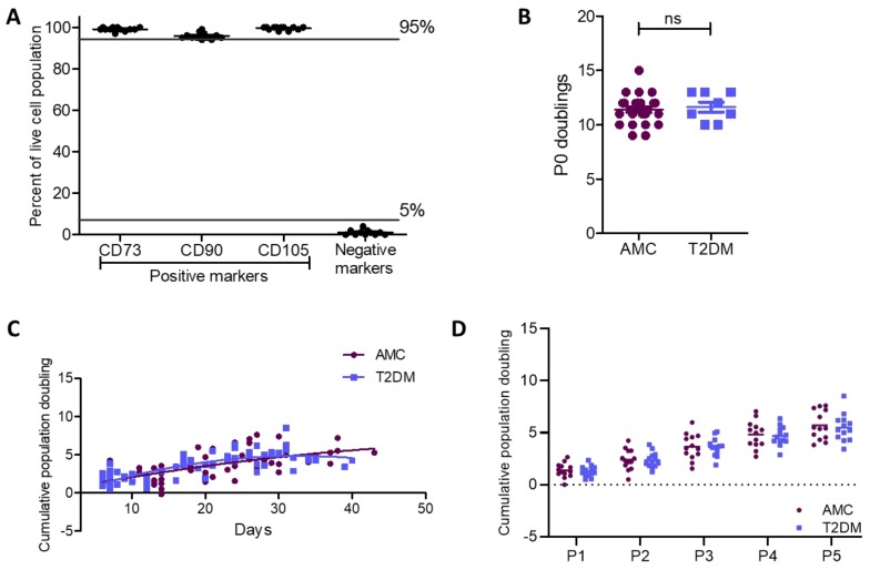
Figure 2.
Cell surface phenotype and proliferative capacity were retained in MSCs from donors with T2DM as compared to AMCs. (A) Flow cytometry confirmed the population of bone marrow-derived primary cells isolated by plastic adherence as being MSCs due to their positive surface marker expression profile (CD73+, CD90+, and CD105+) and absence of CD34, CD45, CD11b or CD14, CD19 or CD79α, and HLA-DR (negative cocktail) expression (n = 12). (B) Population doubling at P0 was unaffected by the diabetic status of the donor (n = 28 for AMC and n = 8 for T2DM cohorts). (C) There was also no difference in doubling capacity between MSCs derived from individuals with T2DM or AMCs when cumulative population doublings per day were compared between the two groups (comparison of second-order polynomial nonlinear regression of the proliferation curve for each cohort of n = 13). (D) A 2-way ANOVA determined that there was no effect of T2DM on proliferative capacity per passage (P1–P5). All data are displayed as mean ± SEM. ns = not significant, i.e., p > 0.05.

