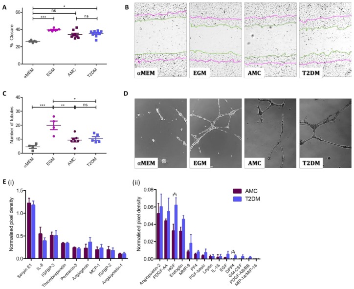
Figure 4.
The presence of T2DM does not influence the capacity of MSC-secreted factors to support angiogenesis. The angiogenic capacity of MSCs to support migration of surrounding cells and to induce tubule formation was evaluated. (A) Quantification of HUVEC cell migration into a scratch in the HUVEC monolayer in the presence of MSC-conditioned medium indicated cells isolated from donors with (n = 10) or without T2DM (n = 8) were capable of stimulating HUVECs to close an in vitro wound. (B) Representative images display a notable decrease in the scratch width as a result of exposure to MSC-conditioned medium regardless of donor cohort. Images obtained by 10× objective as described in methods. Zero h scratch width is overlain onto the photograph taken at 8.5 h, with the 0 h outline traced in purple and the 8.5 h line outlined in green. (C) MSC-conditioned medium was capable of stimulating the formation of endothelial cell tubules regardless of its donor origin (AMC n = 7 and T2DM n = 5). (D) Representative images indicate the presence of long, branching tubules forming complete loops with a comparable morphology in cultures exposed to both types of MSC conditioned medium. Images obtained by 10× objective as described in methods. (E) Angiogenic protein levels in conditioned media at higher concentrations (i) and at lower concentrations (ii). Multiple T-test in R demonstrated increased levels of two secreted proteins (HGF and DPP4, mean ± SEM were 0.0322 ± 0.008 (AMC) and 0.0619 ± 0.009 (T2DM), and −0.002 ± 0.001 (AMC) and 0.003 ± 0.001 (T2DM), respectively), and no significant difference in levels where no statistics summary label is provided on the image (n = 4 per cohort). All data are displayed as mean ± SEM. * = p ≤ 0.05; ** = p ≤ 0.01, *** = p ≤ 0.001, ns = not significant, i.e., p > 0.05.

