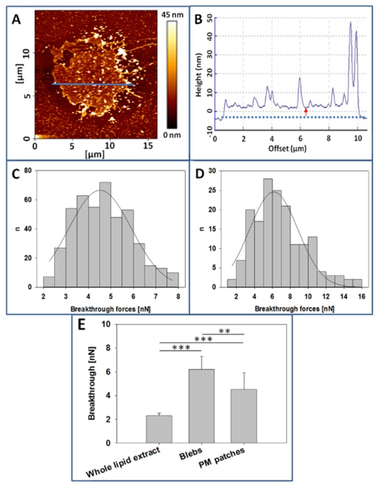
Figure 5.
Atomic Force Microscopy (AFM) measurements. (A), CHO cell plasma membrane patch over polylysine-coated mica. (B), Topographic image of the cross-section indicated by the blue line in 5A. (C), Breakthrough forces distribution of CHO cell plasma membranes (PM) patches. (D). Breakthrough forces distribution of CHO cell blebs. (E). Comparison of breakthrough forces, data extracted from experiments as in C, D. Whole lipid extract breakthrough value was obtained from supported planar bilayers formed with CHO cell lipid extracts. (n= at least 160, value = mean + SD) Statistically significant differences were calculated with ANOVA and Student´s t-test. Significance: (**) p < 0.01; (***) p < 0.001.

