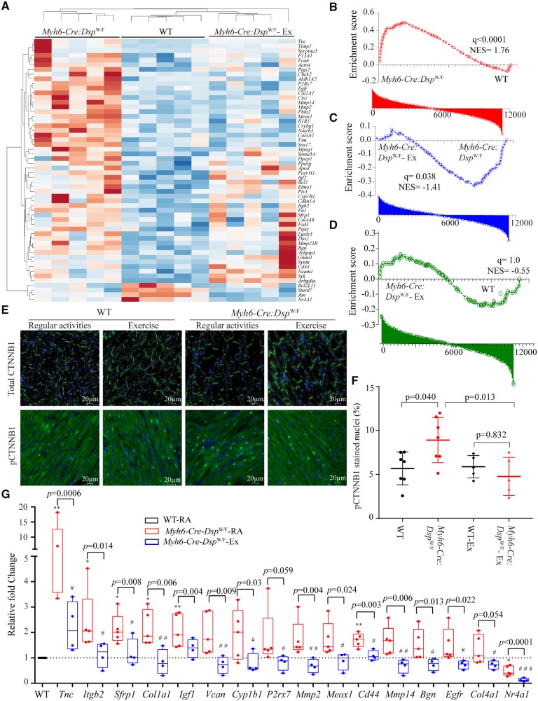
Figure 4.
Rescue of the canonical WNT pathway in Myh6-Cre:DspW/F mice upon treadmill exercise. (A) Heat map of selected known canonical WNT target gene transcripts in Myh6-Cre:DspW/F myocytes and their clustering with the transcripts in WT myocytes with exercise. (B–D) Pairwise gene set enrichment analysis plots and the corresponding q values, showing enrichment of transcript levels of the canonical WNT pathway target genes and the effects of exercise. (B) Myh6-Cre:DspW/F vs. WT myocytes; (C) Myh6-Cre:DspW/F—regular activity vs. Myh6-Cre:DspW/F—exercise; (D) Myh6-Cre:DspW/F—exercise vs. WT myocytes. (E) Representative immunofluorescence panels showing expression and subcellular localization of total and phospho-β catenin (Ser33/37/Thr41) in the experimental groups (N = 7 in WT and Myh6-Cre:DspW/F regular activity groups and N = 5 mice for WT and Myh6-Cre:DspW/F - exercise groups). (F) Quantitative data showing number of nuclei expressing phospho-β catenin. (G) Quantitative PCR depicting transcript levels of selected canonical WNT pathway target genes in myocytes isolated from WT (N = 5, set at 1 for normalization), Myh6-Cre:DspW/F—regular activity (N = 5), and Myh6-Cre:DspW/F—exercise groups (N = 4). The P-values were calculated by ANOVA. *P < 0.05, **P < 0.01 for pairwise comparison between WT and Myh6-Cre:DspW/F—regular activity groups, and #P < 0.05 and ##P < 0.01, respectively, between Myh6-Cre:DspW/F mice in the regular activity and exercise groups.

